[us_page_title align=”center” description=”1″]
Clone JASY1 validated for IHC detection of CD138 plasma cell marker in routine-fixed FFPE tissue sections
CD138/Syndecan-1 antibody
Fig.02 Strong CD138 staining in a basal-cell carcinoma of the skin. Numerous CD138 positive plasma cells reside in the stroma.
Fig.03 Benign dermal nevus: prominent membranous CD138 staining of nomal epidermis. Nevus cells are completely CD138 negative.
Fig.04 Breast ductal carcinoma: Breast cancer (no special type, NST) with strong membranous CD138 staining of tumor cells and intensive CD138 positivity of the tumor stroma.
Fig.05 Tubular breast cancer with strong stromal CD138 immunostaining. Cancer cells are CD138 negative.
Fig.06 Strong membranous CD138 immunostaining of a clear cell kidney cancer.
Fig.07 Focal CD138 positivity in a colon adenoma (mosaic pattern). Multiple CD138 positive plasma cells are present in the stroma.
Fig.08 Colon adenoma with intense predominantly membranous CD138 immunostaining. Numerous CD138 positive plasma cells are located in the stroma.
Fig.09 Strong membranous CD138 immunostaining in a serous carcinoma of the endometrium.
Fig.10 Diffuse membranous CD138 positivity in a serous carcinoma of the endometrium.
Fig.11 CD138 negative serous carcinoma of the endometrium with CD138 positive plasma cells in the tumor stroma.
Fig.12 Weak to moderate membraneous CD138 immunostaining in a hepatocellular carcinoma.
Fig.13 CD138 negative hepatocellular carcinoma. Abundant CD138 positive plasma cells in the tumor stroma.
Fig.14 Strong membraneous CD138 immunostaining of a hepatocellular carcinoma.
Fig.15 Hodgkin lymphoma showing strong CD138 immunostaining of plasma cells.
Fig.16 Strong diffuse, predominantly membraneous CD138 staining of tumor cells in a squamous cell carcinoma of the larynx. In the stroma: numerous CD138 positive plasma cells.
Fig.17 Moderate membraneous CD138 positivity in a bronchiolo-alveolar adenocarcinoma of the lung.
Fig.18 Squamous cell carcinoma of the oral cavity showing strong membraneous CD138 staining of tumor cells. In addition, CD138 positive plasma cells are located in the tumor stroma.
Fig.19 Squamous cell carcinoma of the oral cavity with weak focal cytoplasmic CD138 staining of tumor cells. Strong diffuse CD138 positivity of the tumor stroma.
Fig.20 Endometroid ovarian carcinoma showing moderately intense membranous CD138 immunostaining.
Fig.21 CD138 negative serous ovarian carcinoma showing diffuse CD138 positivity in the tumor stroma.
Fig.22 CD138 negative serous ovarian carcinoma with diffuse CD138 immunostaining in the tumor stroma.
Fig.23 Neuroendocrine tumor of the pancreas with intense membranous CD138 immunostaining.
Fig.24 Papillary renal cell carcinoma with intense membranous CD138 positivity.
Fig.25 Papillary carcinoma of the thyroid with strong membranous CD138 immunostaining in most tumor cells.
Fig.26 Numerous CD138 positive plasma cells are in a CD138 negative papillary carcinoma of the thyroid.
Fig.27 CD138 negative prostate cancer. Adjacent normal prostatic tissue shows CD138 positivity of basal cells.
Fig.28 Renal oncocytoma with membranous and cytoplasmic CD138 positivity.
Fig.29 Squamous cell carcinoma of the skin: Strong membranous CD138 staining.
Fig.30 Strong membranous CD138 positivity in most cells of a thyroid adenoma.
Fig.31 Squamous cell carcinoma of the anal canel showing strong membranous CD138 positivity. Numerous CD138 positive plasma cells are seen in the stroma.
Fig.32 Strong CD138 staining in a squamous cell carcinoma of the anal canel.
CD138/Syndecan-1 antibody

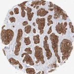
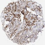
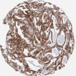
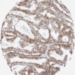
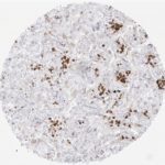
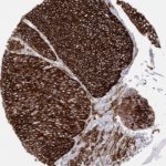
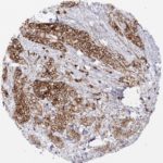
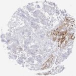
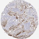
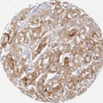
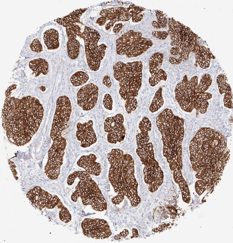
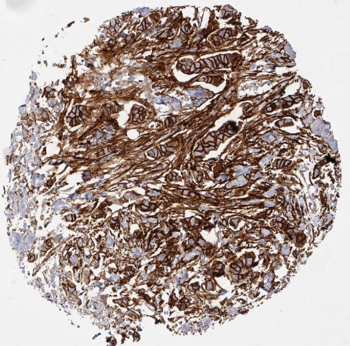
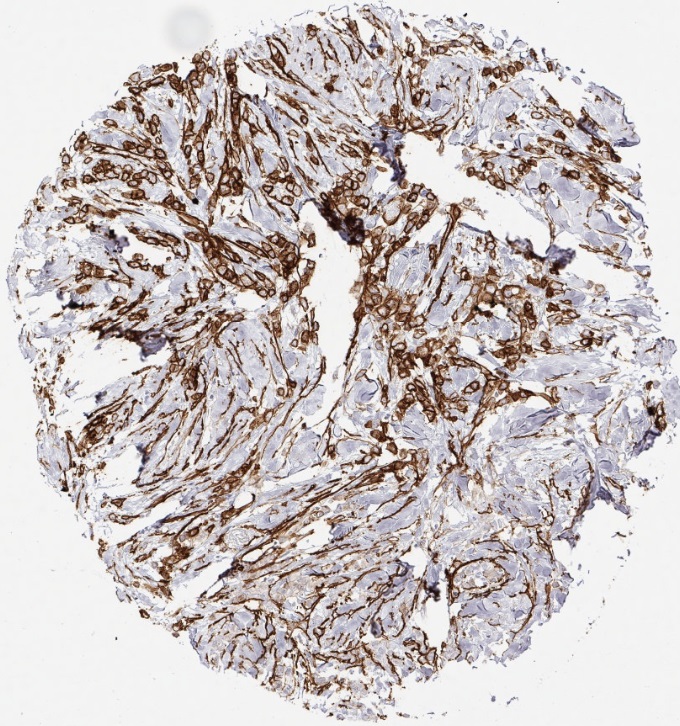
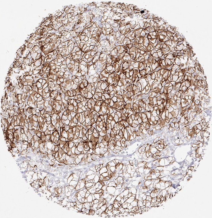
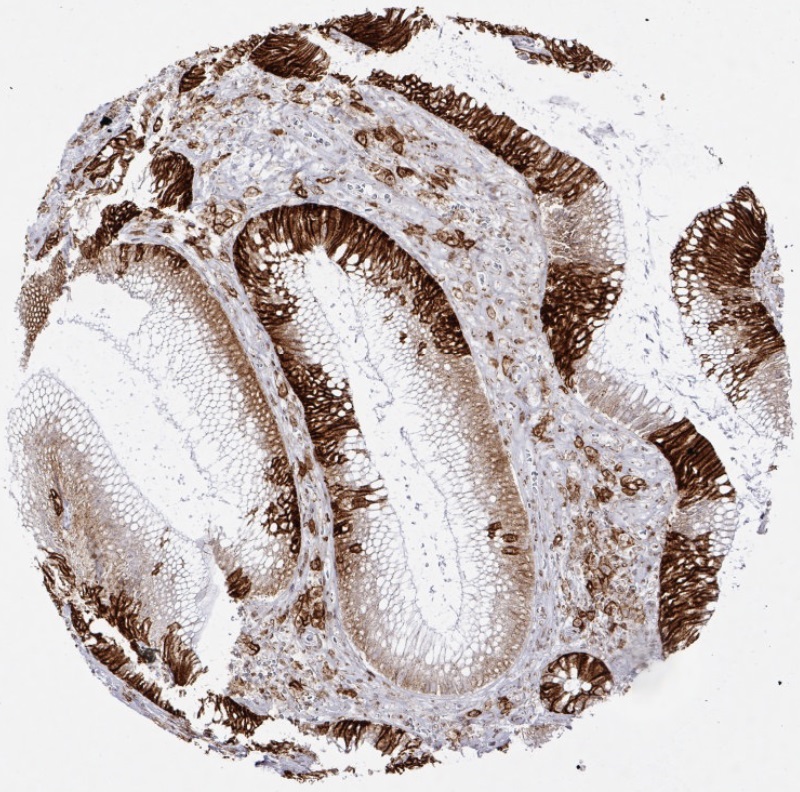
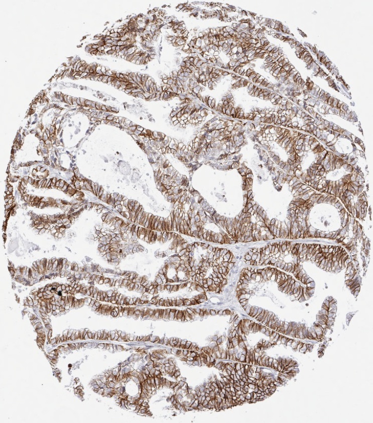
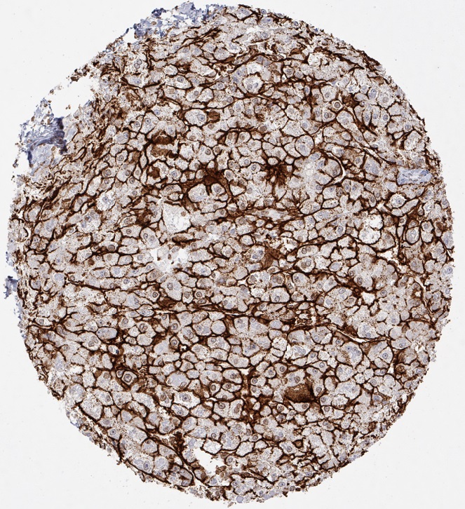
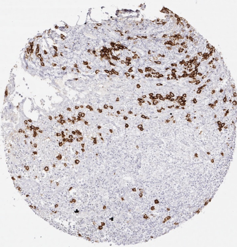
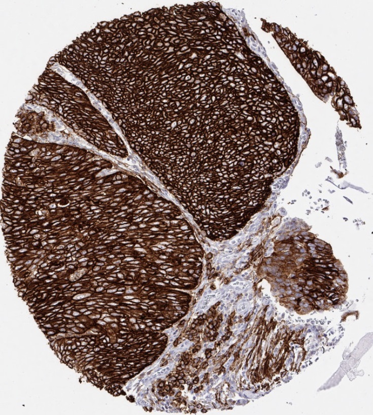
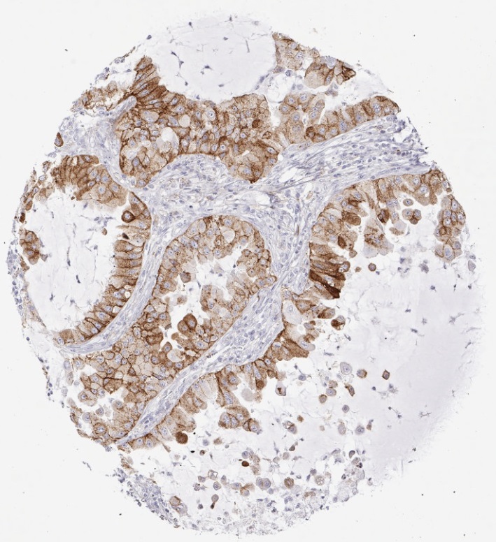
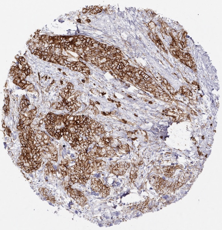
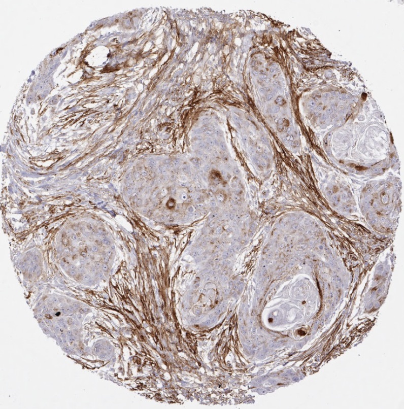
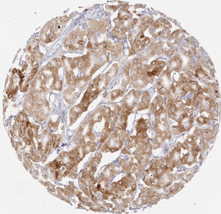
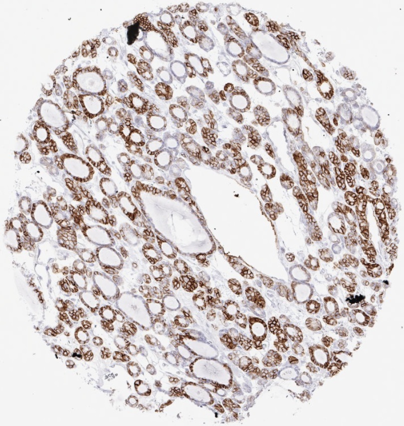
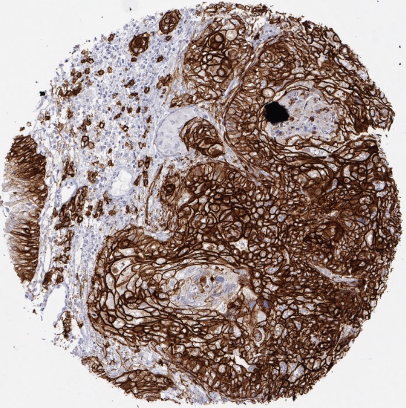
Cluster of differentiation 138 (CD138), also known as Syndecan-1, is a transmembrane glycoprotein, (heparin sulphate proteoglycan) expressed on the surface of plasma cells within the hematopoetic system and on the surface of mature epithelial cells. CD138 is composed of a single chain transmembrane core protein (30,5 KDa, comprising a short cytoplasmic domain, a transmembrane domain, and a long extracellular domain) and five covalently attached glycosaminoglycan.

In diagnostic surgical pathology, antibodies against CD138 are commonly used to identify and quantitate plasma cells. Syndecan-1 (CD138) is a cell surface protein with relevance for cell-cell and cell-matrix interaction. In normal tissues, CD138 is expressed on plasma cells but also in various epithelial cell types. CD138 is also expressed in various cancers. In several tumor types CD138 expression levels were described to be prognostically relevant. CD138 expression in cancer is of potential clinical interest. Antibody-based drugs targeting CD138 in plasmocytomas are being evaluated in clinical trials. In preclinical studies, anti-CD138 antibodies were also effective against triple negative breast cancer and melanoma cells.

1.
Kind S. et al. (2019) A shift from membranous and stromal syndecan-1 (CD138) expression to cytoplasmic CD138 expression is associated with poor prognosis in breast cancer. Mol. Carcinogenesis, 58(12):2306-2315. doi: 10.1002/mc.23119
2.
Kind, S. et al. (2019) Prevalence of Syndecan-1 (CD138) Expression in different kinds of human tumors and normal tissues. Disease Markers, Volume 2019, Article ID 4928315. doi.org/10.1155/2019/4928315

CD138 Antibody Productdetails

Menu