01
Clone-specific evidence
Evidence in which the actual ONCOdianova clone was used and is named — peer-reviewed publications, theses or congress contributions that identify the clone or its catalog number.
Clone-specific

IO·01 Translational immuno-oncology
Study immune-checkpoint pathways and the tumor immune microenvironment in human FFPE tissue.
TIGIT, PVRIG/CD112R, CD73, FOXP3 and CD8 describe complementary dimensions of the tumor immune microenvironment: inhibitory checkpoint expression on lymphocytes, the DNAM-1 axis, adenosine-mediated immune regulation, regulatory T cells and cytotoxic T-cell infiltration. ONCOdianova develops mouse monoclonal antibodies for the immunohistochemical study of these targets in human formalin-fixed, paraffin-embedded (FFPE) tissue — in conventional brightfield IHC and, where documented, in multiplex immunohistochemistry. This page describes how each target is being investigated in immune-checkpoint research, translational immuno-oncology and treatment-response biomarker research, and it separates that target-level literature from the evidence that exists for each ONCOdianova clone.
For Research Use Only. Not for use in diagnostic procedures.
IO·02 Evidence model
Every statement on this page is assigned to one of three evidence levels. The levels are not interchangeable: what has been shown for a target in independent research is not thereby shown for an ONCOdianova clone.
Evidence in which the actual ONCOdianova clone was used and is named — peer-reviewed publications, theses or congress contributions that identify the clone or its catalog number.
Clone-specific
ONCOdianova evidence: datasheets, FFPE-IHC protocols, tissue figures and multiplex images produced by ONCOdianova for the clone in question.
ONCOdianova validation
Independent research on the target in immuno-oncology, tumor-microenvironment or treatment-response biomarker research, obtained with other reagents or methods.
Target-level
Target-level translational evidence does not imply clinical validation of the ONCOdianova clone.Target-level findings describe what is known about a molecule; they do not describe the analytical or clinical performance of a specific antibody clone. ONCOdianova antibodies are research reagents. For Research Use Only. Not for use in diagnostic procedures.
IO·03 TIGIT — clones TG1 and TG2
TIGIT (T-cell immunoreceptor with Ig and ITIM domains) is an inhibitory immune-checkpoint receptor of the poliovirus-receptor family. It is expressed on subsets of T lymphocytes and NK cells, competes with the co-stimulatory receptor CD226 for the ligands CD155 and CD112, and is a therapeutically investigated target. Tissue-based TIGIT expression has been investigated by brightfield and multiplex immunohistochemistry on human FFPE tissue.
Blessin et al. analyzed TIGIT expression across 1,778 tumors representing 86 tumor entities, together with normal and inflammatory tissues, using clone TG1 with Western blotting, ELISA and pre-absorption reported as specificity assessments.[1] More recent research has investigated TIGIT expression in relation to the tumor immune contexture and to treatment response: in melanoma, TIGIT expression within the tumor microenvironment was analyzed together with immune-cell infiltrates and immunohistochemistry, and a high TIGIT-positive lymphocyte score was associated with longer progression-free survival under anti-PD-1 immunotherapy in that cohort.[2]In breast cancer, the CD155–TIGIT/CD96/CD226 axis was evaluated by immunohistochemistry on tumor cells and tumor-infiltrating lymphocytes and related to prognosis.[3] These are findings on the target obtained with the reagents of the respective studies.
Clone TG1 is identified by clone or catalog number in twelve peer-reviewed publications, including the systematic study of 1,778 tumors across 86 entities.[1] TG1 can be used to study TIGIT-positive lymphocytes in human FFPE tissue by brightfield and fluorescence multiplex IHC.
Clone TG2 is documented by its own datasheet with tonsil as positive control and a membranous lymphocyte staining pattern; compared with TG1 it shows higher affinity for TIGIT, stronger tonsil staining intensity and higher dilutability. TG2 has not itself been studied in treatment-response research; no such claim is made.
Independent studies have investigated tissue TIGIT expression in relation to immune contexture, prognosis and anti-PD-1 therapy.[2, 3] These findings concern the target and the reagents of those studies; they are not evidence for clone TG1 or TG2.
Related research context: CD8 — cytotoxic T-cell infiltration · FOXP3 — regulatory T cells · PVRIG/CD112R — DNAM-1 axis.
IO·04 PVRIG / CD112R — clone R12
Terminology. PVRIG is the receptor and is identical to CD112R. Its ligand is PVRL2, also called CD112 or nectin-2. CD112R (the receptor) and CD112 (the ligand) are different molecules.
PVRIG/CD112R was identified as an inhibitory checkpoint for human T cells that binds PVRL2/CD112 and competes with the co-stimulatory receptor DNAM-1 (CD226) for this ligand.[4]PVRIG and PVRL2 are induced in cancer and inhibit CD8-positive T-cell function.[5]COM701 is an investigational therapeutic anti-PVRIG antibody; it has been studied alone and in combination with nivolumab, and with a TIGIT antibody and nivolumab, in registered phase 1 and phase 1/2 trials in advanced solid tumors.[7, 8] Tissue PVRIG expression is therefore a target of translational interest for immunohistochemistry on human FFPE tumor tissue.
In a translational analysis presented at SITC 2023, pretreatment (n = 28) and on-treatment (n = 21) biopsies from patients with platinum-resistant ovarian cancer treated with COM701 plus nivolumab, with or without the TIGIT antibody BMS-986207 (NCT03667716 and NCT04570839), were stained by immunohistochemistry with anti-PD-L1, anti-CD8, anti-PVRL2 and anti-PVRIG. In this preliminary analysis the authors report clinical responses independent of baseline PD-L1, CD8 and PVRIG expression, a correlation of higher baseline PVRL2 expression with clinical benefit, and an increase in CD8-positive cells in the on-treatment biopsies of 8 of 13 patients with paired samples.[9]Immunohistochemistry for PVRIG and its ligand PVRL2 in FFPE biopsies is thus part of the biomarker work that accompanies clinical PVRIG blockade.
In colorectal cancer, CD112R/PVRIG and PD-1 were analyzed on cytotoxic T lymphocytes by fluorescence multiplex immunohistochemistry on tissue microarrays and large sections of 523 tumors with follow-up, using clone R12.[6]
Clone R12 (DIA-R12) is named in a peer-reviewed fluorescence multiplex IHC study of CD8, PD-1, CD112R and Ki67 in 523 colorectal cancers.[6] R12 can be used to study CD112R/PVRIG-positive lymphocyte subsets in human FFPE tissue, alone or in multiplex panels.
ONCOdianova documents CD112R × CD8 and CD112R × FOXP3 fluorescence multiplex staining of human tonsil with clone R12, together with brightfield IHC figures and a documented FFPE protocol.
PVRIG biology,[4, 5] the registered COM701 trials[7, 8] and the immunohistochemical biomarker analysis of trial biopsies[9] establish target-level translational relevance for tissue PVRIG research. Clone-specific evidence for R12 is listed on the product page.
Related research context: TIGIT — the second checkpoint of the CD155/CD112 ligand family · CD73 — adenosine-mediated immune regulation.
IO·05 CD73 / NT5E — clone KK3
CD73 (ecto-5′-nucleotidase, NT5E) participates in extracellular adenosine generation by converting AMP to adenosine, a pathway that contributes to immune regulation in the tumor microenvironment. CD73 is under therapeutic investigation. Coveler et al. (2024) evaluated CD73 expression in exploratory biomarker analyses within a randomised study of oleclumab-based combinations in metastatic pancreatic cancer. This provides a clinical-study example of CD73 biomarker research alongside investigation of adenosine-targeted treatment approaches.[10]
Clone KK3 is a research reagent for tissue-based CD73 studies in this context; associations between CD73 expression patterns and treatment response are investigated in exploratory, study-specific analyses.
Clone KK3 (DIA-KK3) is documented by its datasheet with retrieval, detection-system-dependent dilutions, tonsil as control, a membranous staining pattern and clone-labelled figures of tonsil, placenta and tumor tissues. KK3 can be used to study CD73 expression on tumor and stromal cells in human FFPE tissue.
CD73 expression has been evaluated in exploratory biomarker analyses within clinical studies of anti-CD73 therapy, providing clinical-study examples of CD73 translational biomarker research.[10]
Published studies provide translational target-level context for CD73 research in the tumor microenvironment and in adenosine-targeted therapy studies. For Research Use Only. Not for use in diagnostic procedures.
Related research context: PVRIG/CD112R — DNAM-1 axis.
IO·06 FOXP3 — clone FX3
FOXP3 is the transcription factor used to study regulatory T-cell populations in the tumor microenvironment. FOXP3-positive populations have been included in tissue-based and multiplex analyses of patients receiving checkpoint immunotherapy: in the PEMBRO-RT trial in non-small cell lung cancer, tumor biopsies taken at baseline and during treatment with pembrolizumab, alone or combined with stereotactic body radiotherapy, were stained by multiplex immunofluorescence for CD3, CD8, CD20, CD103 and FOXP3. In that exploratory study, lymphocyte infiltration in general, rather than a specific lymphocyte subset, was associated with response.[11]
The biological meaning of FOXP3-positive infiltrates is context-dependent and differs between cancer types, compartments and treatments; no single direction of association with response is generalized here.
Clone FX3 (DIA-FX3) is named in a peer-reviewed study that used automated multiplex immunofluorescence on human FFPE tissue.[12] FX3 can be used to study FOXP3-positive, nuclear-staining lymphocytes in human FFPE tumors by brightfield and multiplex IHC.
ONCOdianova documents FOXP3 × CD112R/PVRIG multicolor immunofluorescence of human tonsil with clones FX3 and R12, together with brightfield FOXP3 figures and a documented FFPE protocol.
FOXP3-positive populations are analyzed in tissue-based and multiplex studies of checkpoint immunotherapy.[11] These findings concern the target and the reagents of those studies; they are not evidence for clone FX3.
Related research context: TIGIT — checkpoint expression on T-cell subsets · CD8 — cytotoxic T-cell infiltration.
IO·07 CD8 — clone TC8
CD8 is a central marker for cytotoxic T-cell infiltration and one of the most widely used immunohistochemical read-outs of the tumor immune microenvironment. CD8 IHC has been used in paired pretreatment and on-treatment biopsies in immunotherapy trials: across early-phase trials of novel immunotherapeutic agents, CD8-positive T cells were stained immunohistochemically on paraffin sections and digitally enumerated in paired biopsies from 155 patients (training set) and 221 patients (validation set). A composite of the on-treatment fold change and the on-treatment density of CD8-positive T cells was associated with progression-free survival and discriminated clinically active from inactive compounds.[13] In the PEMBRO-RT trial, CD8 was part of the multiplex panel applied to baseline and on-treatment biopsies.[11]
Beyond paired biopsies, CD8 IHC is used to classify the spatial immune phenotype of tumors. In a tumor-agnostic analysis of 2,023 patients from 14 phase I–III clinical trials, pathologists classified tumors as CD8-inflamed, CD8-excluded or CD8-desert on the basis of CD8 IHC staining in the epithelial and stromal areas of the tumor; this IHC-based spatial classification served as the reference for a transcriptome-based classifier of the CD8 immunophenotype.[14]
These associations were obtained with the assays of the respective studies and describe how CD8 density is being used in treatment-response biomarker research.
Clone TC8 (DIA-TC8) was used in five peer-reviewed studies on several thousand human FFPE tumors, in automated brightfield IHC with digital quantification of CD8-positive cells per mm² and in Opal multiplex immunofluorescence.[15, 16] TC8 can be used to study CD8-positive tumor-infiltrating lymphocytes and their density in human FFPE tissue.
ONCOdianova documents CD8 × CD112R/PVRIG fluorescence multiplex staining of human tonsil with clones TC8 and R12, together with a large brightfield gallery and a documented FFPE protocol.
Changes in CD8-positive T-cell density in paired biopsies have been associated with clinical outcome in published trial analyses,[11, 13] and CD8 IHC has been used to classify tumors from 14 clinical trials into inflamed, excluded and desert immune phenotypes.[14] Clone TC8 is a research reagent for tissue-based CD8 studies in this context.
Related research context: TIGIT — checkpoint expression on CD8-positive T cells · FOXP3 — regulatory T cells · PVRIG/CD112R — checkpoint on cytotoxic T cells.
IO·08 Portfolio
The combination shown here represents a research framework, not a validated diagnostic or treatment-selection panel. Each antibody is a research reagent with its own, separately documented evidence. For Research Use Only. Not for use in diagnostic procedures.
IO·09 Multiplex
Tumor immune biology is multi-marker and spatial: which cells express a checkpoint, where they sit relative to tumor cells and to each other, and how the populations change under treatment. Multiplex immunohistochemistry addresses these questions on a single FFPE section. The ONCOdianova portfolio includes antibodies used for conventional and, where documented, for multiplex IHC — clone R12 in a peer-reviewed four-marker panel,[6] clone TC8 in Opal multiplex immunofluorescence,[16]clone FX3 in automated multiplex immunofluorescence,[12] and clone TG1 in fluorescence multiplex IHC among its clone-specific publications.
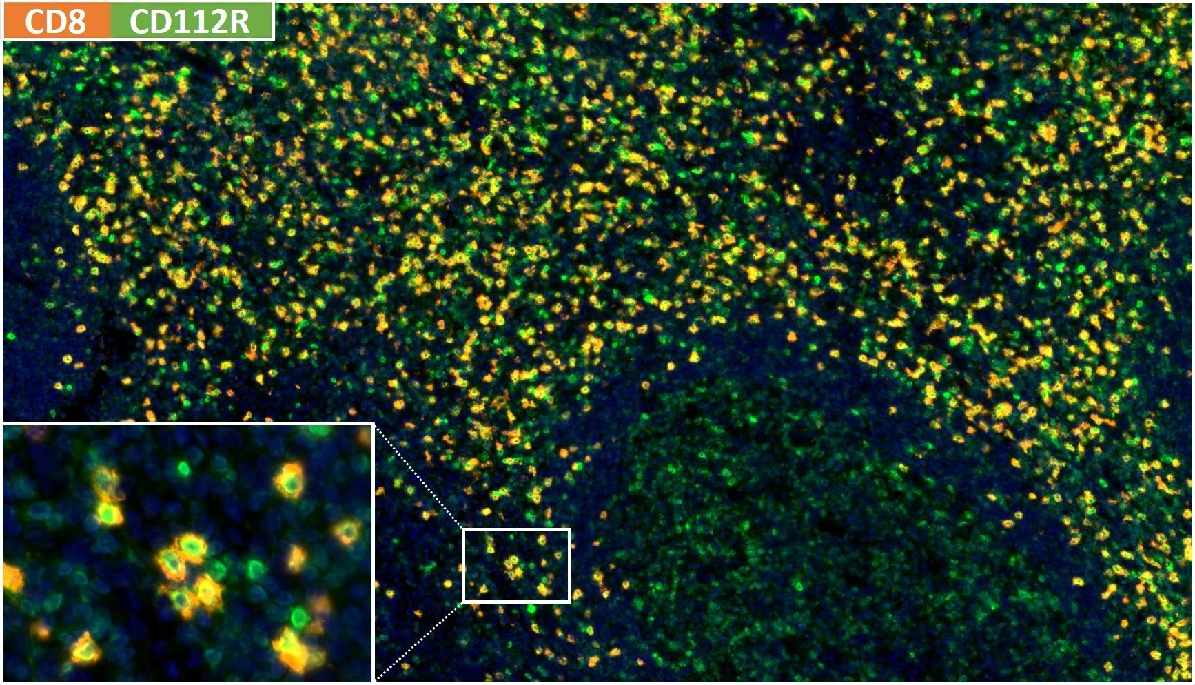
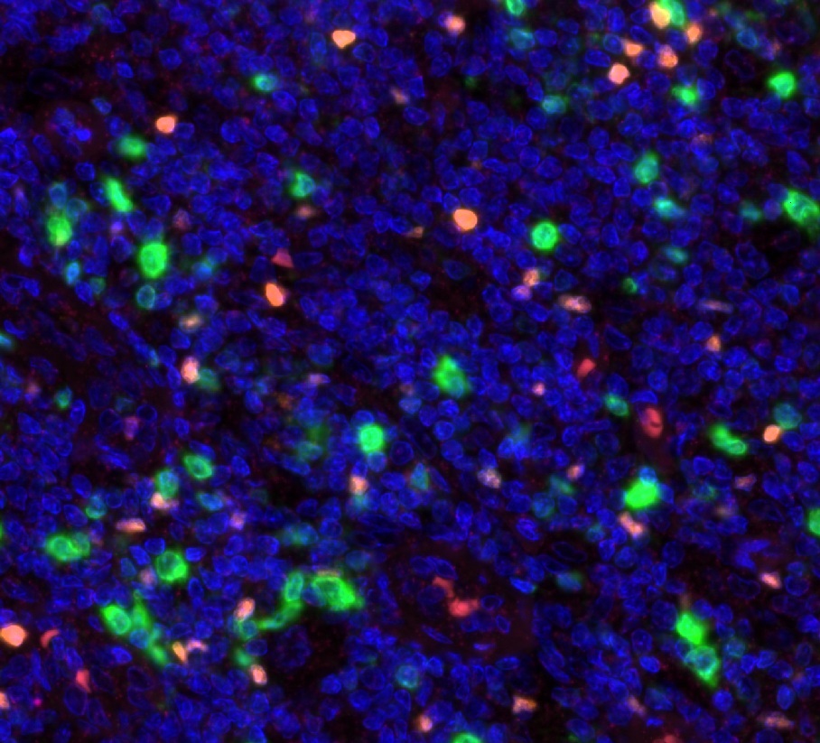
Both images are original ONCOdianova material with their legends. Multiplex use of a clone is stated only where a clone-specific publication or an ONCOdianova figure documents it; combinations of markers are a research framework, not a validated panel.
IO·10 References
“Clone-specific” marks sources in which the named ONCOdianova clone was used; “Target-level” marks independent research on the target obtained with other reagents. Target-level translational evidence does not imply clinical validation of the ONCOdianova clone.
Product data, protocols, galleries and clone-specific publications are documented on each product page. For Research Use Only. Not for use in diagnostic procedures.