What does anti-FOXP3 clone FX3 detect?
FX3 is a mouse monoclonal antibody (IgG2a/κ) raised against a recombinant peptide of human FOXP3 (forkhead box protein P3; gene FOXP3). It is offered as a research reagent for immunohistochemistry on human formalin-fixed, paraffin-embedded (FFPE) tissue, in brightfield IHC and in fluorescence multiplex IHC, with a nuclear staining pattern.
Is FX3 documented for FFPE immunohistochemistry?
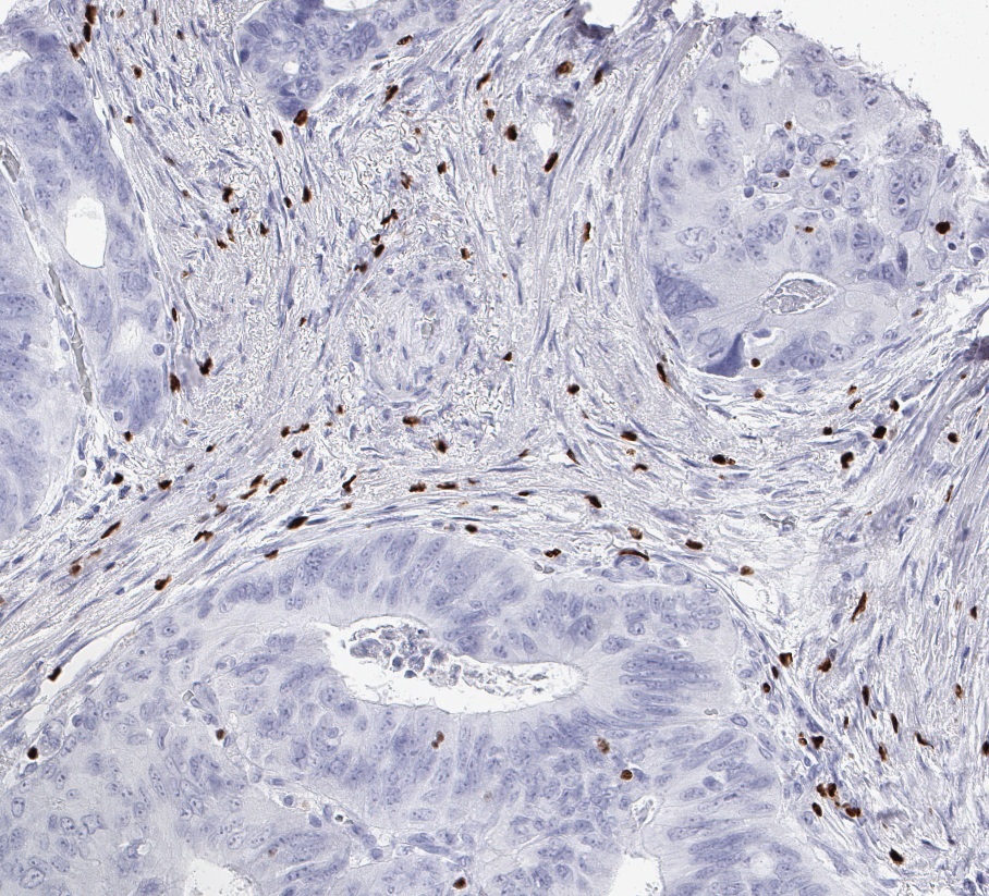
Yes. Clone FX3 was developed for the detection of FOXP3 in routine formalin-fixed, paraffin-embedded tissue specimens; the datasheet gives a starting range of 1:100 to 1:200 (IHC-P). ONCOdianova figures show nuclear FOXP3 staining in tonsil, colorectal adenocarcinoma, high-grade serous ovarian carcinoma and further solid cancers.
Are there peer-reviewed publications using clone FX3?
Yes. Picard et al., Acta Neuropathologica 2023 (PMID 37656187) used DIA-FX3 at 1:200 in an automated six-marker multiplex immunofluorescence panel (Opal Polaris 7-colour kit) on human FFPE sections of pilocytic astrocytoma.
Is FX3 validated for fluorescence multiplex IHC?
Yes. Clone FX3 has been developed and validated for fluorescence multiplex IHC studies of FOXP3 expression in human tissues (ONCOdianova product information and datasheet), and the gallery shows FOXP3 × CD112R/PVRIG multicolor immunofluorescence of normal tonsil. Published application: Picard et al. 2023 used DIA-FX3 in an automated Opal multiplex panel. Every new panel should be verified for tissue, antibody order, retrieval, dilution and detection.
Which dilution is a suitable starting point for FX3?
The datasheet recommends 1:100 to 1:200 (IHC-P) on FFPE sections; the ONCOdianova manual protocol uses 1:100. The published multiplex study used 1:200 within an Opal panel – a study-specific condition. The optimal dilution should be determined by the user for each tissue, fixation, platform and detection system.
Which epitope retrieval does FX3 require?
Heat-induced epitope retrieval is required. The datasheet recommends an autoclave at 121 °C for 5 minutes in Tris-EDTA-citrate buffer, pH 7.8 (TEC buffer).
Which positive control and staining pattern are documented for FX3?
Human tonsil, with a nuclear staining pattern of FOXP3-positive lymphocytes. The datasheet names tonsil as the positive-control tissue and “nuclear” as visualization; the gallery includes a tonsil overview and FOXP3 × CD112R/PVRIG multiplex images of normal tonsil.
How can FOXP3-positive cells be evaluated in tissue research?
FX3 visualizes nuclear FOXP3 protein in FFPE tissue. Depending on the research question, FOXP3-positive cells can be evaluated together with morphology, tissue compartment and complementary immune markers.
What is the documented validation scope of clone FX3?
Clone FX3 was developed for the detection of FOXP3 in routine FFPE tissue, has been characterized on hundreds of tissues and validated for fluorescence multiplex IHC studies of FOXP3 expression in human tissues; it detects FOXP3-positive tumour-infiltrating lymphocytes in FFPE human tumours (ONCOdianova product information). Published clone-specific application: automated multiplex immunofluorescence on human FFPE tissue with DIA-FX3 at 1:200 (Picard et al. 2023).
Can FX3 be used on automated staining platforms?
Yes. The datasheet states that the antibody is suited for immunohistochemical staining on automated platforms, and the ONCOdianova product information names it applicable for automated staining procedures. Picard et al. 2023 performed their multiplex staining with DIA-FX3 on the LabSat™ automated instrument.