PAX8

328,00 plus VAT

#DIA-PAX8-OD
Anti-PX8 (Hu) from Mouse (clone JAX8) – 100 µl
Category:

Description

at.No. DIA-PX8-OD
Specificity PAX8
Species Reactivity Human
Host Species Mouse
Isotype IgG2a
Clone JAX8
Application
Immunohistochemistry (IHC),
Formalin-fixed Paraffin-embedded sections (FFPE)
Western Blot
Conjugation unconjugated
Dilution IHC 1:100-1:200
Format
Liquid
Antibody purified (from culture supernatant)
Quantity 100µl
Control tissue Ovarian (non-mucinous), Thyroid or Renal Cell Carcinoma
Presentation Tris pH 7.3-7.7, with 1% BSA, <0.1% NaN3
Intended Use Research Use Only
 Manufacturer / Brand ONCOdianova
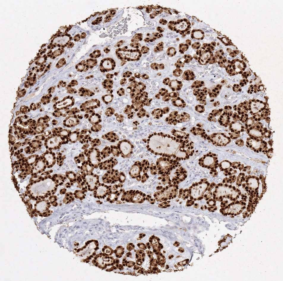
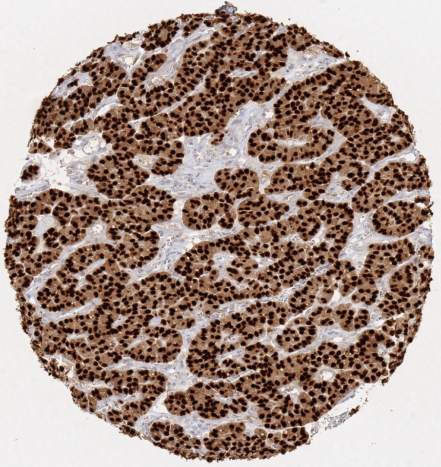
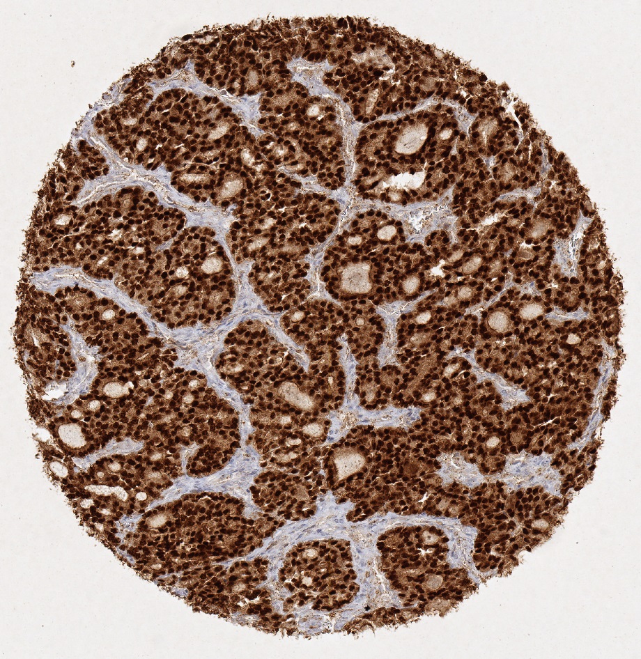
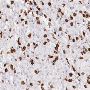
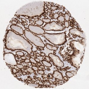
PAX8 IHC-Gallery
IHC-figures and legends on PAX8 detection in different tumors.

Reactivity

Mouse monoclonal anti-PAX8 antibody clone JAX8 is suitable for the immunohistological detection of PAX8 in routine-fixed paraffin embedded tissue sections.

PAX8 is a member of the paired box (PAX) family of transcription factors involved the regulation of early development of the thyroid gland, kidney, and Müllerian tract. PAX8 plays a central role for the expression of thyroid-specific genes and thus, in development of thyroid follicular cells. Mutations in the PAX-8 gene are linked to thyroid follicular carcinomas. In the develop-ing kidney PAX8 is important for renal vesicle formation.

PAX8 is highly expressed in several neoplasms: Epithelial tumors of the thyroid and parathyroid glands, kidney, thymus, pan-creatic neuroendocrine tumors and female genital tract. Follicular and papillary thyroid carcinoma are almost always PAX8 positive (while medullary thyroid carcinoma is negative, but anaplastic carcinoma is positive in most cases). PAX8 is also found in almost all cases of endometrial carcinoma and ovarian serous, endometrioid, transitional and clear cell carcinoma. Moreover, PAX8 is found in most cases of renal cell carcinoma (all types) and in oncocytoma as well as in thymic tumours. Difefferent reports have shown that adenocarcinomas of lung and breast are negative for PAX8-expression. Also, PAX-8 is not found in the epithelial cells of the breast, lung, mesothelium, stomach, colon, pancreas.

PAX 8 is a useful IHC marker with a wide range of diagnostic applications and appears to be the most specific and sensitive marker for renal cell carcinoma and ovarian non-mucinous carcinoma.

References

References

  1. Marques AR et al. (2002) Expression of PAX8 PPAR gamma 1 rearrangements on both follicular thyroid carcinomas and andenomas. J Clin Endocrinol Metab. 87: 3947-3952
  2. Nikiforova MN et al. (2002) PAX8-PPARgamma rearrangement in thyroid tumors: RT-PCR and immunohistochemi-cal analyses. Am J Surg Pathol. 26: 1016-1023
  3. Zhang P, et al. (2006) Immunohistochemical analysis of thyroid‐specific transcription factors in thyroid tumors. Pathol Int. 56: 240-245
  4. Bowen NJ, et al. (2007) Emerging roles for PAX8 in ovarian cancer and endosalpingeal development. Gynecol On-col. 104: 331-337
  5. Nonaka, D et al. (2008) Diagnostic utility of thyroid transcription factors Pax8 and TTF-2 (Fox E1) in thyroid epithelial neoplasms. Mod Pathol. 21: 192-200
  6. Nonaka D et al. (2008) Expression of pax8 as a useful marker in distinguishing ovarian carcinomas from mammary carcinomas. Am J Surg Pathol. 32: 1566-157
  7. Tong GX et al. (2009) Expression of PAX8 in normal and neoplastic renal tissues: an immunohistochemical study. Mod Pathol. 22: 1218-1227
  8. Laury AR et al. (2011) A comprehensive analysis of PAX8 expression in human epithelial tumors. Am J Surg Pathol. 35: 816-826
  9. Ozcan A et al. (2011) PAX 8 expression in non-neoplastic tissues, primary tumors, and metastatic tumors: a compre-hensive immunohistochemical study. Mod Pathol.; 24: 751-764

You may also like…

Menu