Clone JAP16 validated for IHC detection of p16 (INK4A) tumor supressor in routine-fixed FFPE tissue sections
Fig.01 Immunohistochemical p16 positivity in a Brenner tumor of the ovary.
Fig.02 Mucinous ovarian carcinoma with p16 positivity in tumor stroma. The neoplastic urothelium shows no staining.
Fig.03 Endometrioid ovarian carcinoma with diffuse p16 positivity.
Fig.04 Endometrioid ovarian carcinoma with focal nuclear p16 positivity. Most of the neoplastic epithelium is p16 negative. Only single cells and small groups of cells show strong nuclear staining (mosaic pattern).
Fig.05 Small proportions of a serous ovarian carcinoma with strong p16 positivity.
Fig.06 Strong immunohistochemical p16 positivity in a serous ovarian carcinoma.
Fig.07 Strong p16 positivity in superficial foci of a serous ovarian carcinoma. Next to it not neoplastic epithelium (p16 negative).
Fig.08 p16 negative serous ovarian carcinoma (stained with p16 clone JAP16).
Fig.09 Strong immunohistochemical p16 positivity in a serous ovarian carcinoma.
Fig.10 Intense p16 positivity in a medullary breast carcinoma. The staining is predominantly cytoplasmic, but also nuclear.
Fig.11 Strong p16 positivity in a medullary breast carcinoma.
Fig.12 Tubular breast carcinoma (p16 negative) with p16 staining of spindle-shaped stromal cells.
Fig.13 Strong nuclear p16 staining in the mesenchymal component of a mammary phyllodes tumor. The epithelium is p16 negative.
Fig.14 Very intense nuclear p16 staining in the mesenchymal component of a phyllodes tumor of the breast. The epithelium shows a weak, also nuclear stressed p16 staining of a few single cells.
Fig.15 Endometrioid endometrial carcinoma. Heterogeneous p16 staining with exclusive p16 positivity of plateepithelial differentiated tumor portions.
Fig.16 Endometrioid endometrial carcinoma with p16 "mosaic-type" staining. The neoplastic epithelium is predominantly p16 negative. However, single cells and small groups of cells show a marked predominantly cytoplasmic staining.
Fig.17 Endometrioid endometrial carcinoma with diffuse, strong p16 staining.
p16 antibody
Fig.18 Serous endometrial carcinoma with strong p16 staining. The stroma is completely p16 negative.
Fig.19 Strong cytoplasmic and nuclear p16 staining in a cervical adenocarcinoma.
Fig.20 Strong cytoplasmic and nuclear p16 staining in a cervical adenocarcinoma.
Fig.21 Moderately strong cytoplasmic and nuclear p16 staining in a cervical adenocarcinoma.
Fig.22 Squamous cell carcinoma of the vagina: Strong cytoplasmic and nuclear immunohistochemical p16 positivity. Squamous cell carcinoma of the vagina: Strong cytoplasmic and nuclear immunohistochemical p16 positivity.
Fig.23 Strong, predominantly cytoplasmic p16 positivity in a gastric carcinoma of the intestinal type.
Fig.24 Strong p16 positivity in adenocarcinoma of the papilla Vateri. The staining is cytoplasmic and nuclear.
Fig.25 Adenocarcinoma of the papilla Vateri. Moderately strong, quite predominant nuclear p16 positivity.
Fig.26 Strong p16 positivity in a breast cancer "no special type (NST)"
Fig.27 Chromophobic renal cell carcinoma: Colorful picture of p16 stains. Single cells show repeatedly low, moderately strong or strong p16 positivity.
Fig.28 Oncocytoma of a kidney. The tumor itself is p16 negative. However, some small vessels are clearly positive p16.
Fig.29 Strong cytoplasmic and nuclear p16 staining in a muscle invasive urothelial carcinoma.
Fig.30 Immunohistochemical p16 staining of a colonic adenoma (low grade).
Fig.31 Strong cytoplasmic and nuclear p16 staining in a muscle invasive urothelial carcinoma.
Fig.32 Immunohistochemical p16 staining of a colonic adenoma (low grade). The neoplastic epithelium is predominantly p16 negative. However, single cells and small groups of cells show p16 staining, predominantly nuclear.
Fig.33 Pheochromocytoma. Some cells show clear cytoplasmic as well as nuclear p16 staining.
Fig.34 Colon adenoma (low grade). The neoplastic epithelium is mostly p16 negative. However, single cells and small groups of cells show strong, predominantly nuclear, p16 positivity.
Fig.35 Strong diffuse p16 staining in an anal carcinoma.
Fig.36 Moderately strong, nuclear-enhanced p16 staining of a gastrointestinal stromal tumor.
Fig.37 Neuroendocrine tumor (carcinoid); About 50% of the tumor cells show a moderately strong, nuclear stressed p16 staining.
Fig.38 Strong p16 staining (nuclear stressed) in a basal cell carcinoma.
Fig.39 Basalioma with strong nuclear p16 positivity of single cells.
Fig.40 p16 negative anaplastic thyroid carcinoma with strong p16 staining of small vessels.
Fig.41 Leiomyosarcoma spot Fa4b
Fig.42 Leiomyosarcoma: Strong, nuclear-stressed p16 positivity in all tumor cells.
Fig.43 Thymoma with nuclear p16 positivity.
p16 antibody
Fig.44 Oral carcinoma: p16 positive.
Fig.45 Oral carcinoma: p16 negative (stained with clone JAP16).
p16 antibody
Fig.46 Oral carcinoma: p16 staining is restricted to the peripheral cells of invasive tumor cell aggregates.
Fig.47 Strongly p16 positive (nuclear and cytoplasmic) laryngeal carcinoma. In between, is an island of normal epithelium isolated from the staining.
p16 antibody
Fig.48 Heterogeneous p16 staining in a non-Hodgkin's lymphoma.
Fig.49 Selective stain of Reed-Sternberg and Hodgkin's cells in a Hodgkin's lymphoma.
Fig.50 Diffuse cytoplasmic p16 staining in a granular cell tumor.
Fig.51 Diffuse vigorous p16 staining of vessels in an angiosarcoma.
Fig.52 p16 staining of atypical lipocytes in a highly differentiated liposarcoma.
Fig.53 Strong, nuclear-enhanced p16 positivity in a dedifferentiated portion of a liposarcoma.
Fig.54 Liposarcoma: Intense nuclear p16 staining.
Fig.55 Malignant mesothelioma with diffuse strong p16 positivity of tumor cells.
Fig.56 Malignant mesothelioma with clear staining of spindle-shaped stromal cells. The epithelioid tumor parts are p16 negative.
Fig.57 Intense p16 staining in a Merkel cell carcinoma of the skin.
p16 antibody
Fig.58 Merkel cell carcinoma of the skin: Strong nuclear p16 positivity.
Fig.59 Intense cytoplasmic, but also nuclear p16 positivity in a small cell neuroendocrine carcinoma of the prostate.
Fig.60 Small cell bladder neuroendocrine carcinoma. Intense, nuclear-enhanced p16 staining.

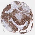
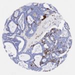
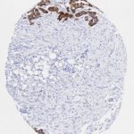
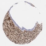
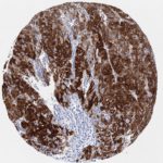
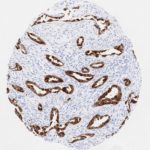
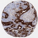
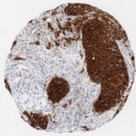
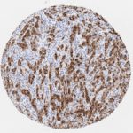
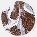
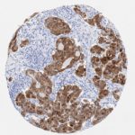
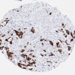
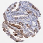
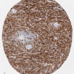
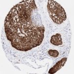
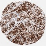
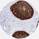
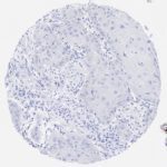
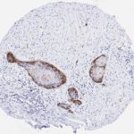
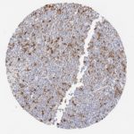
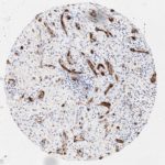
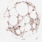
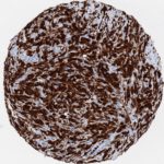
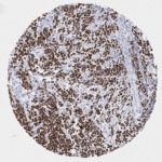
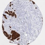
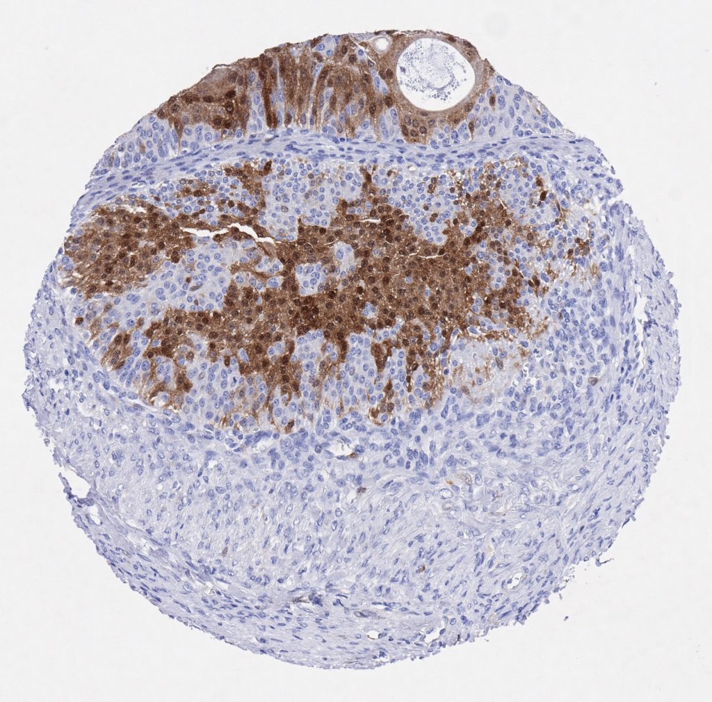
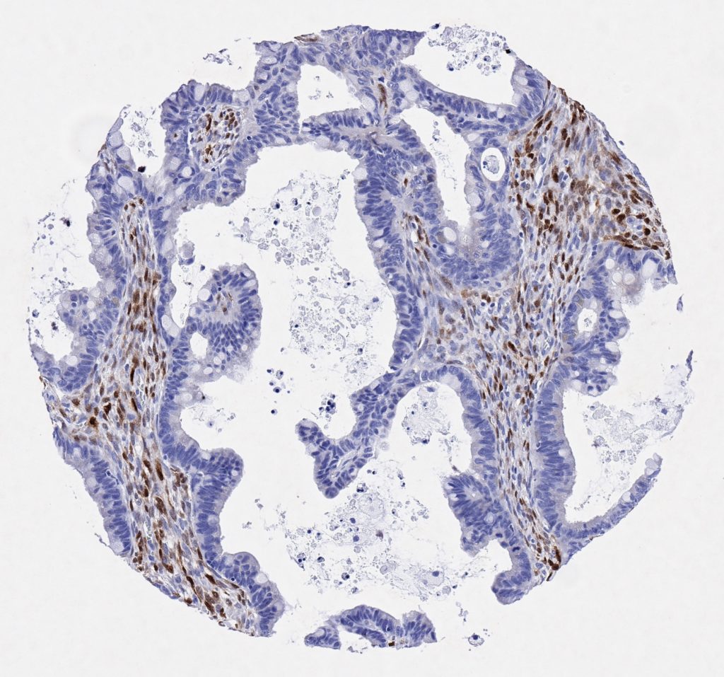
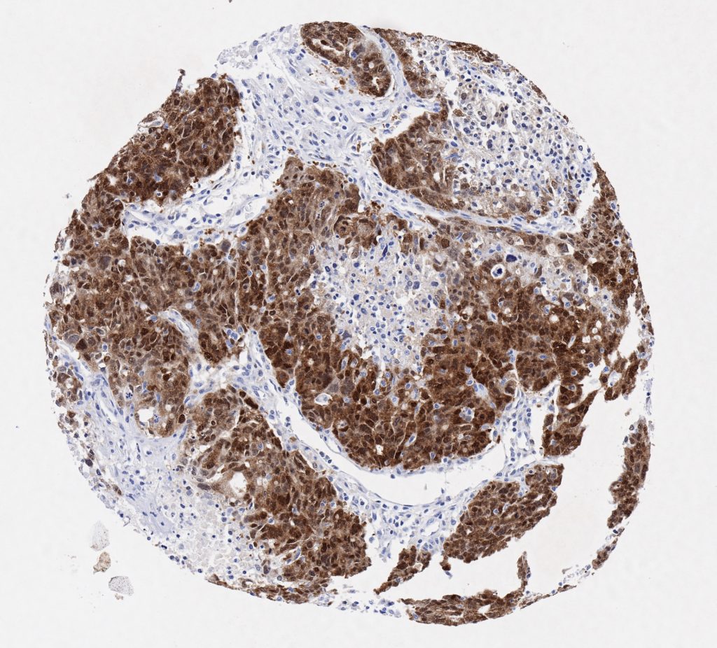
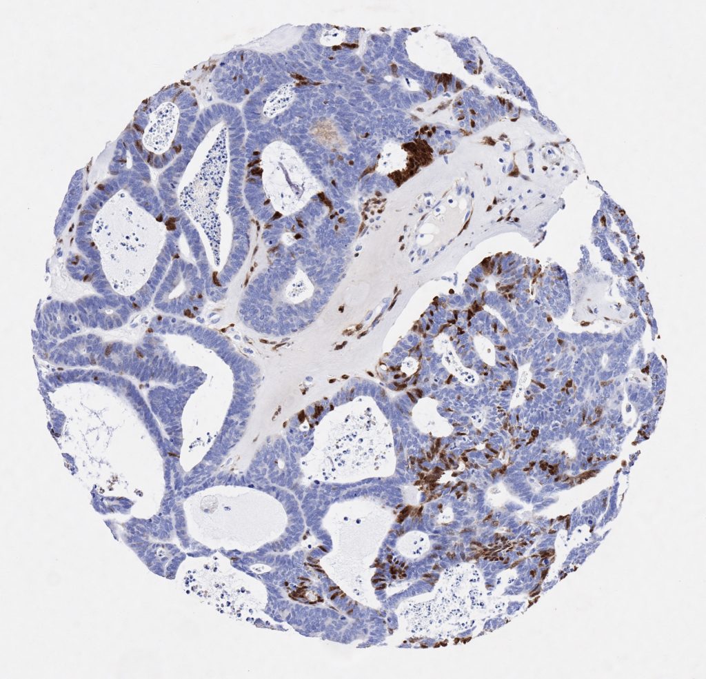
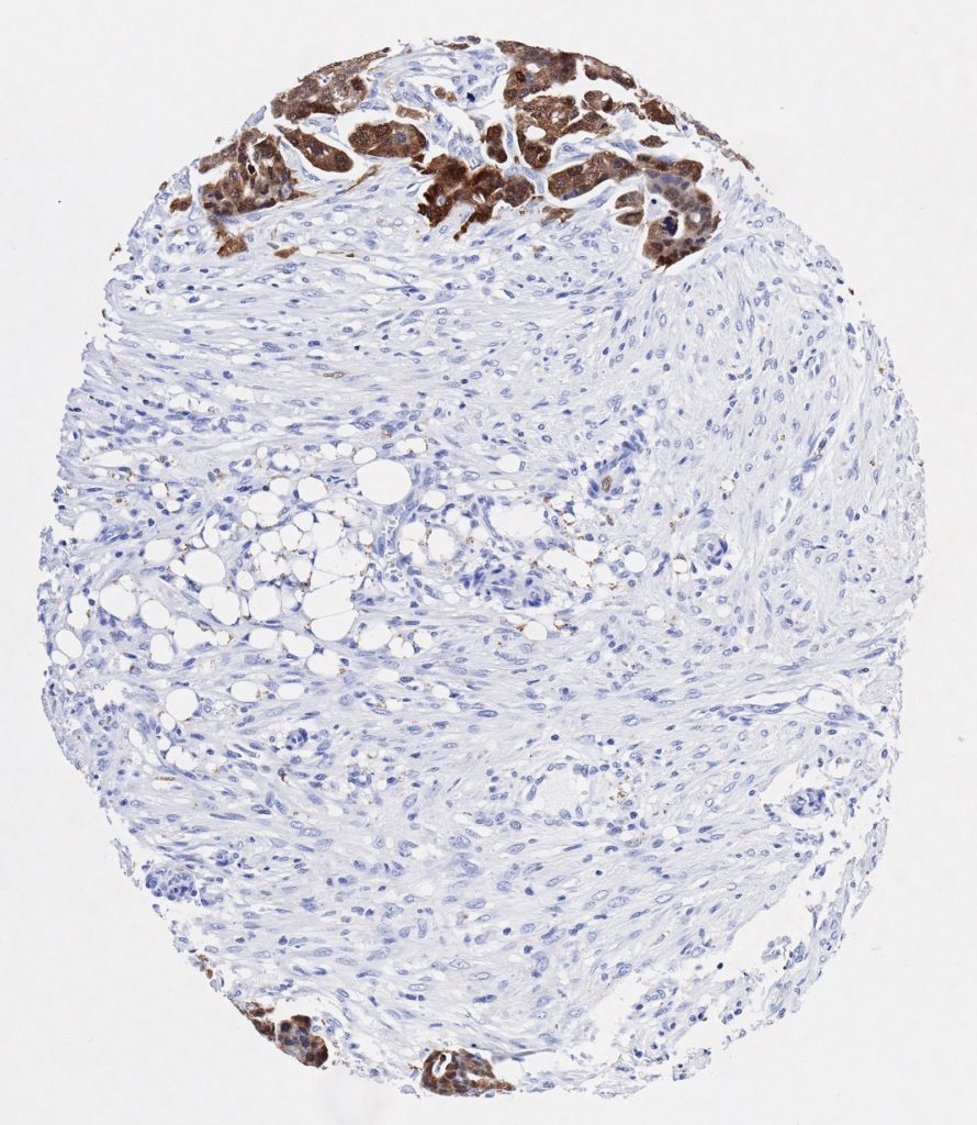
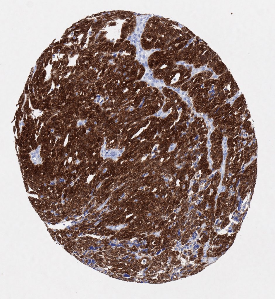
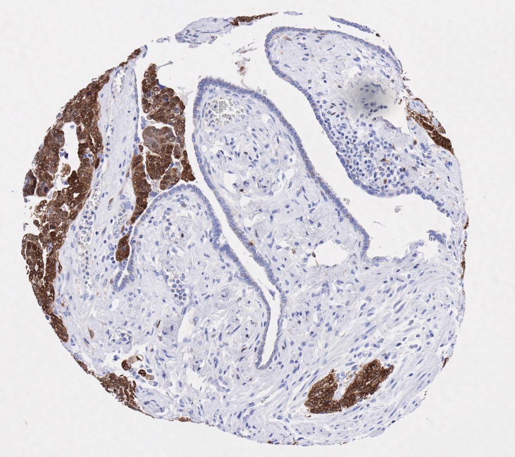
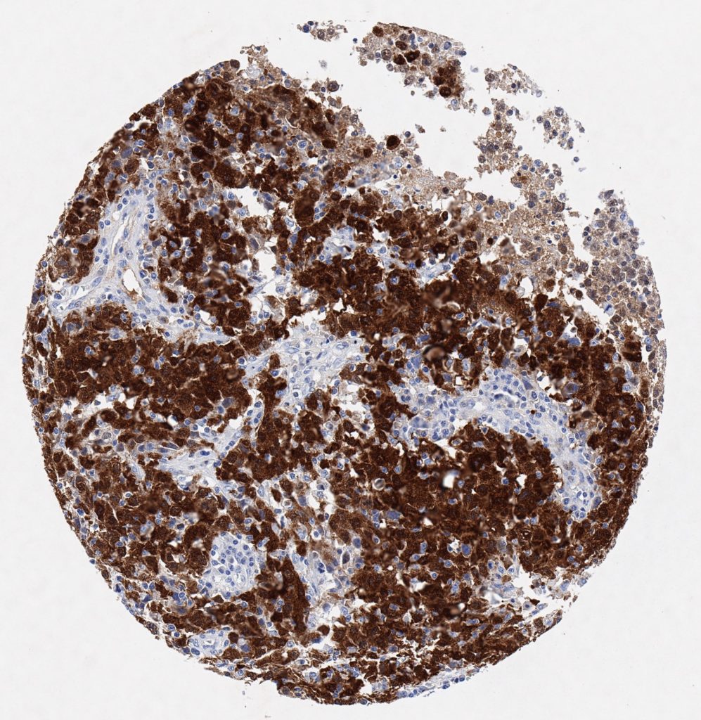
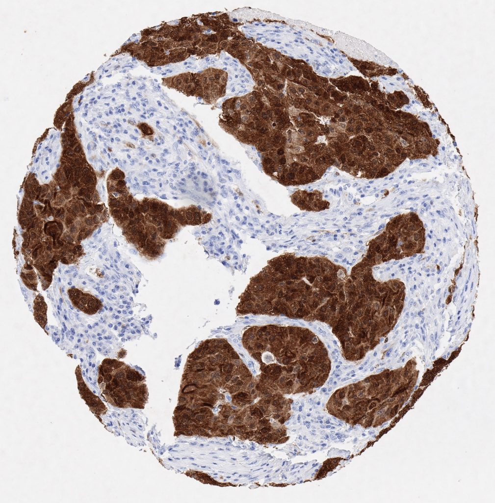
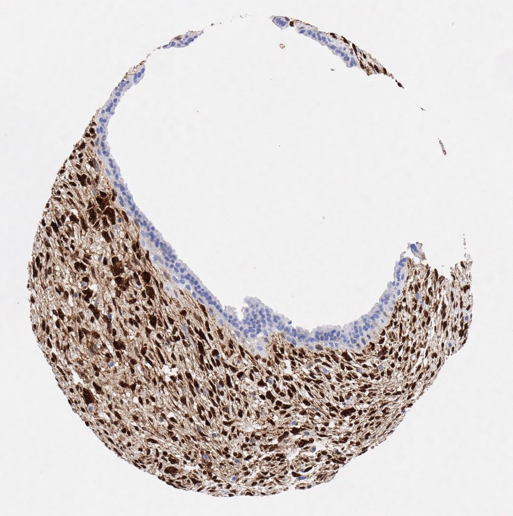
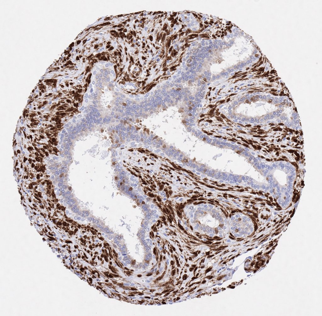
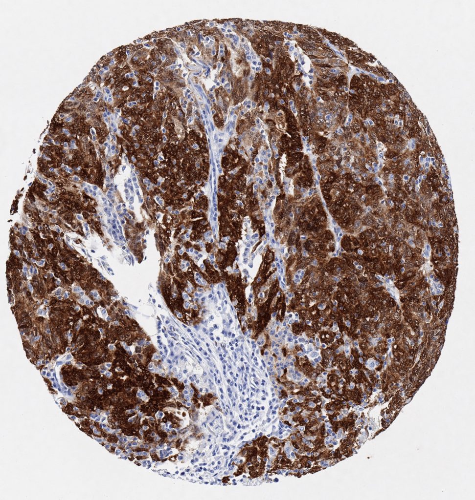
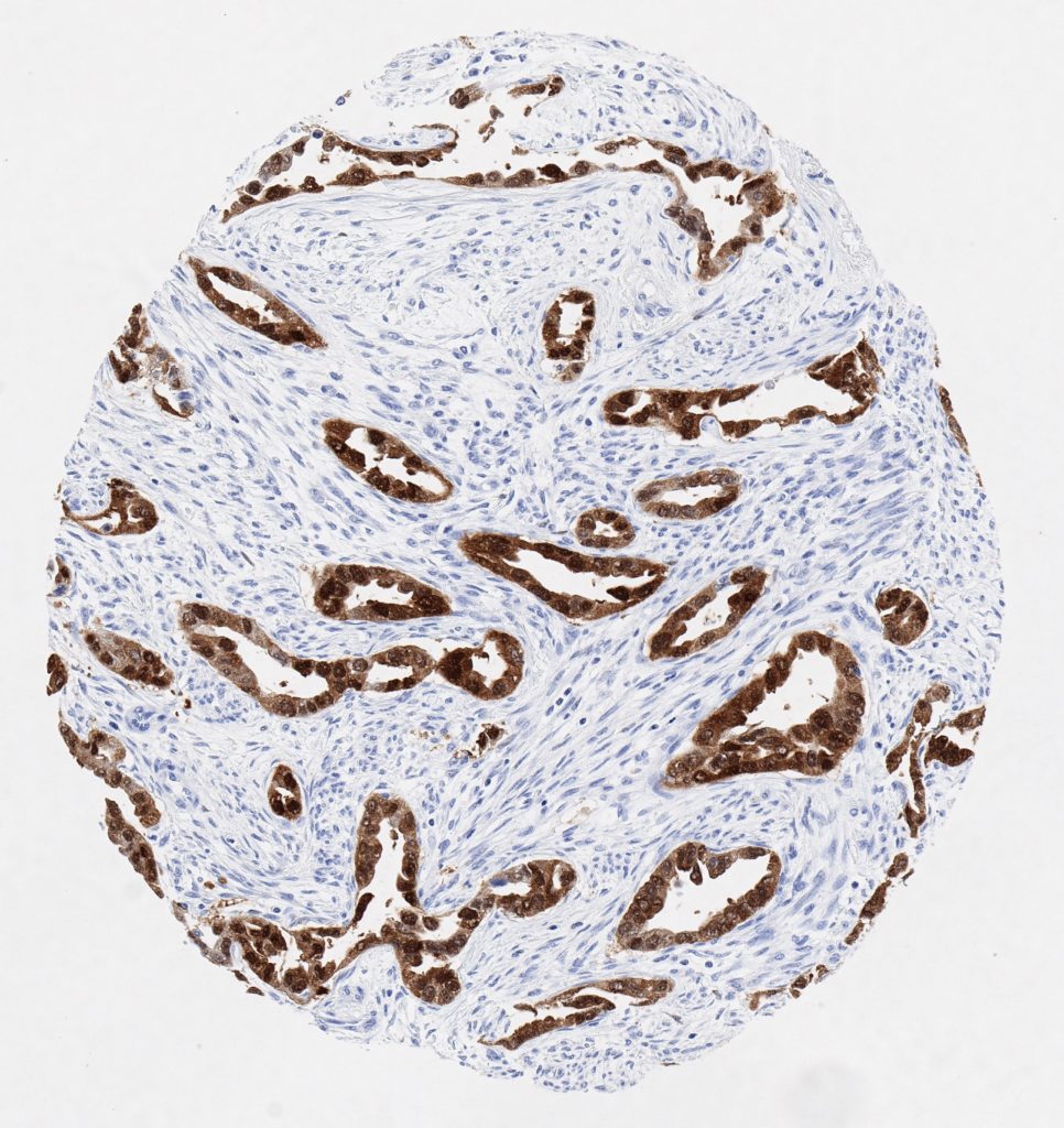
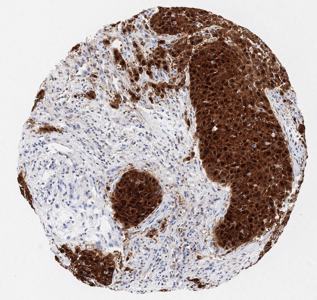
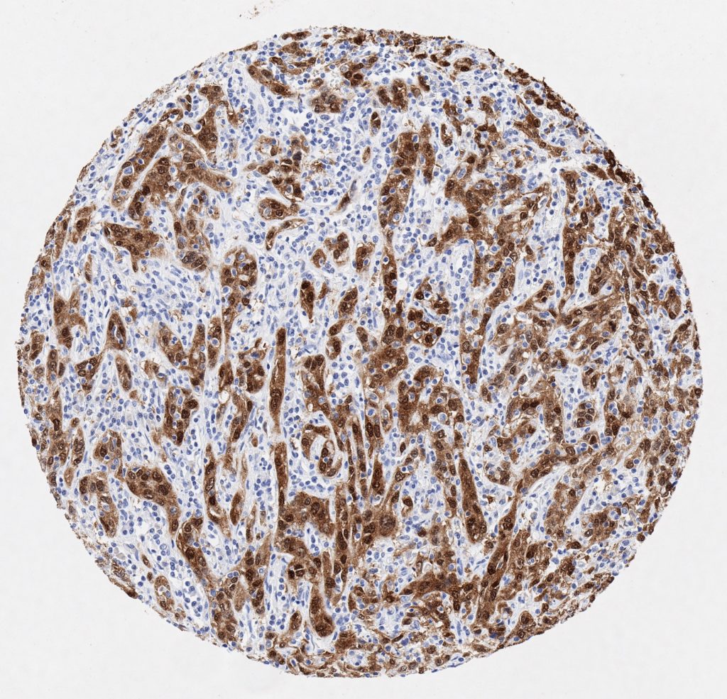
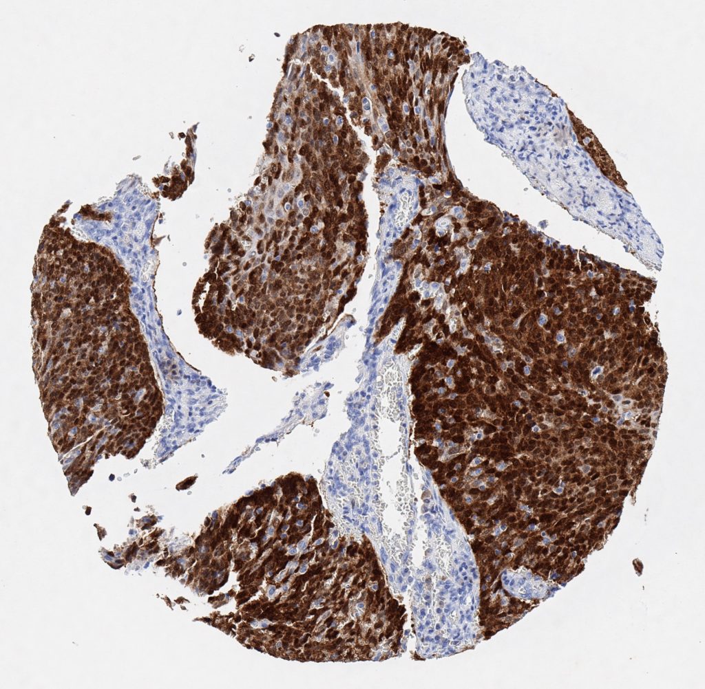
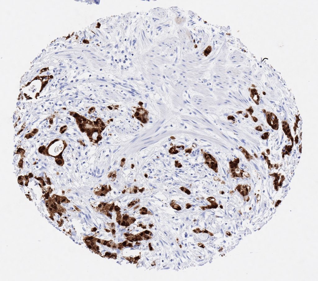
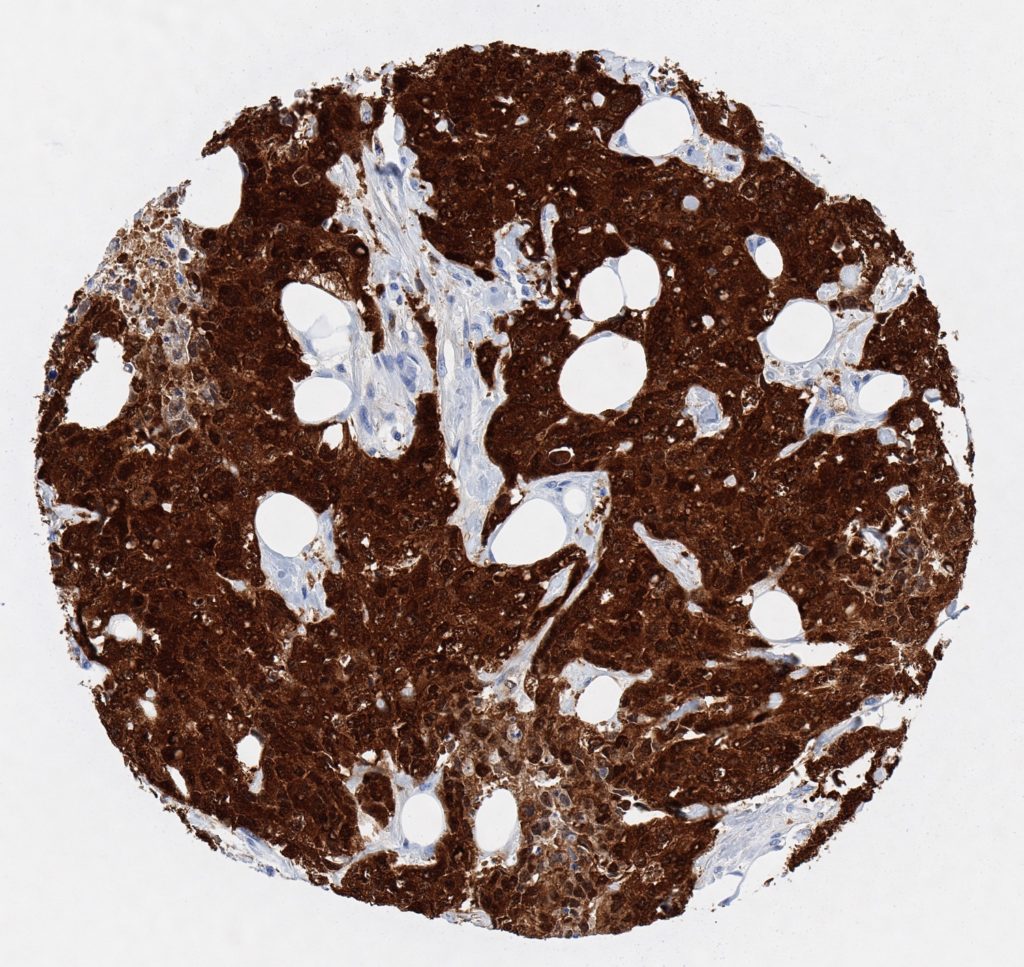
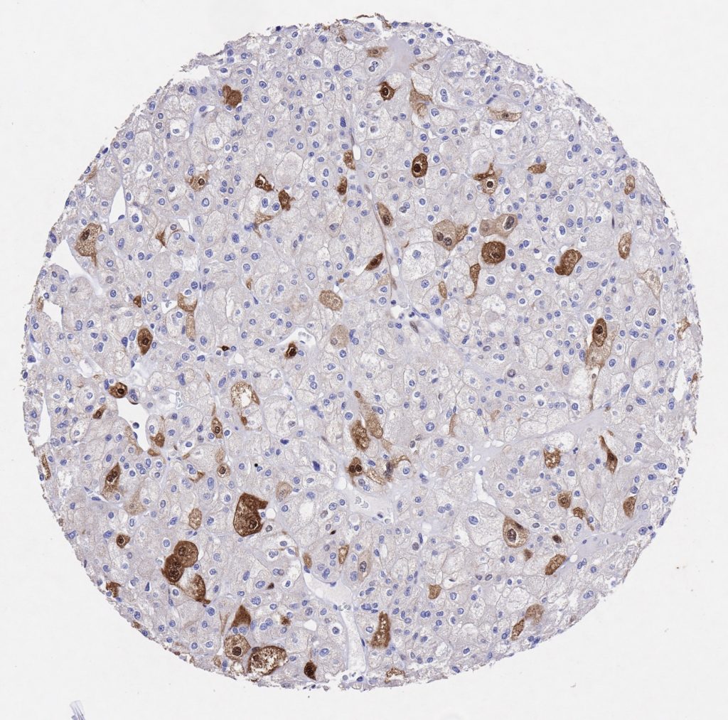
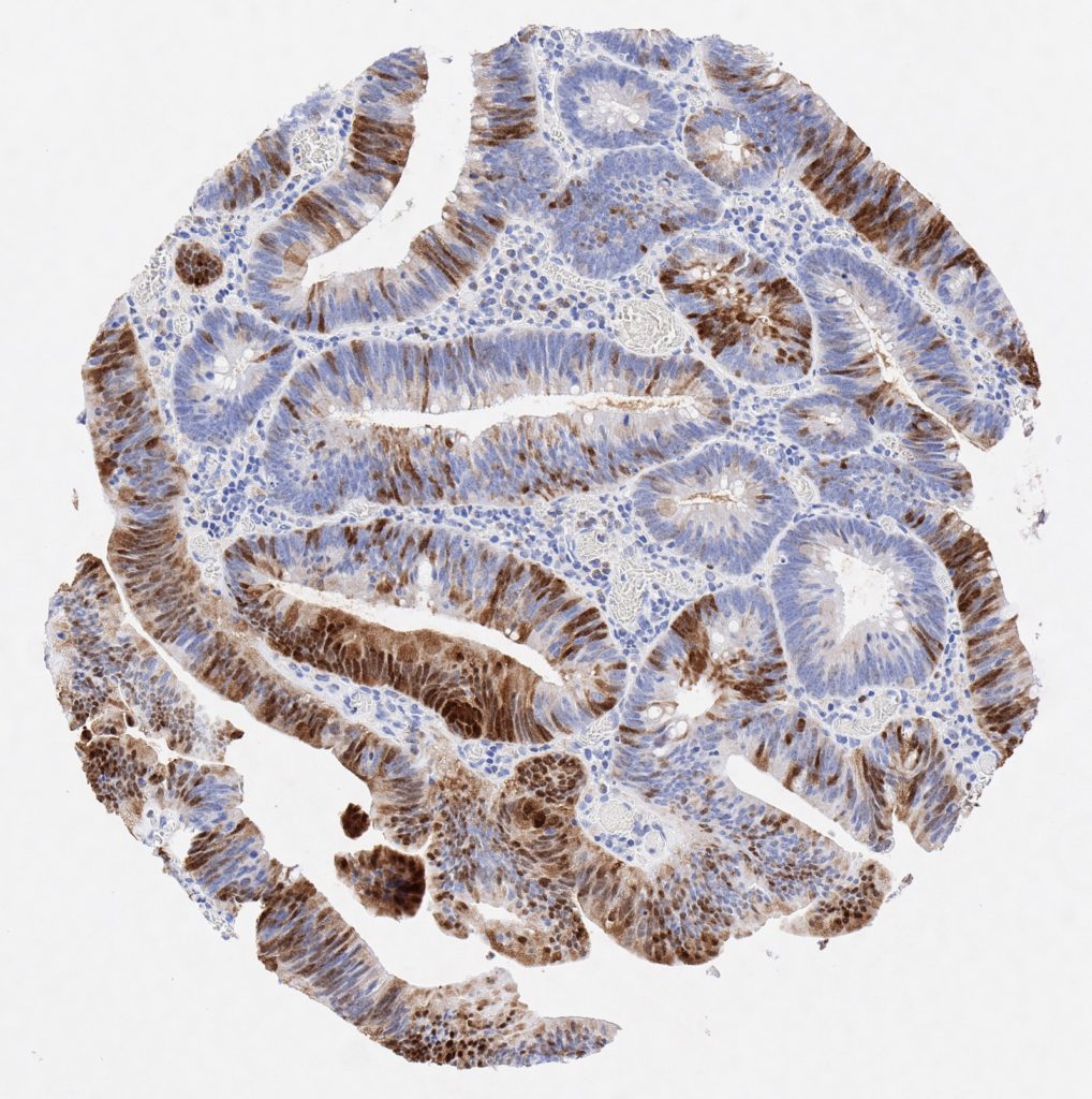
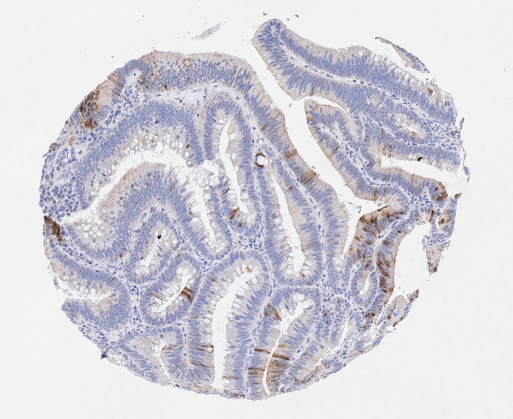
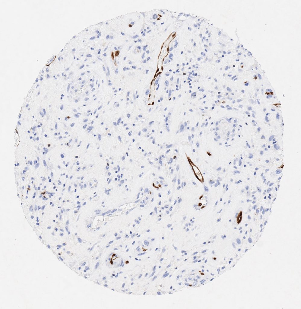
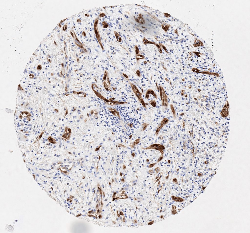
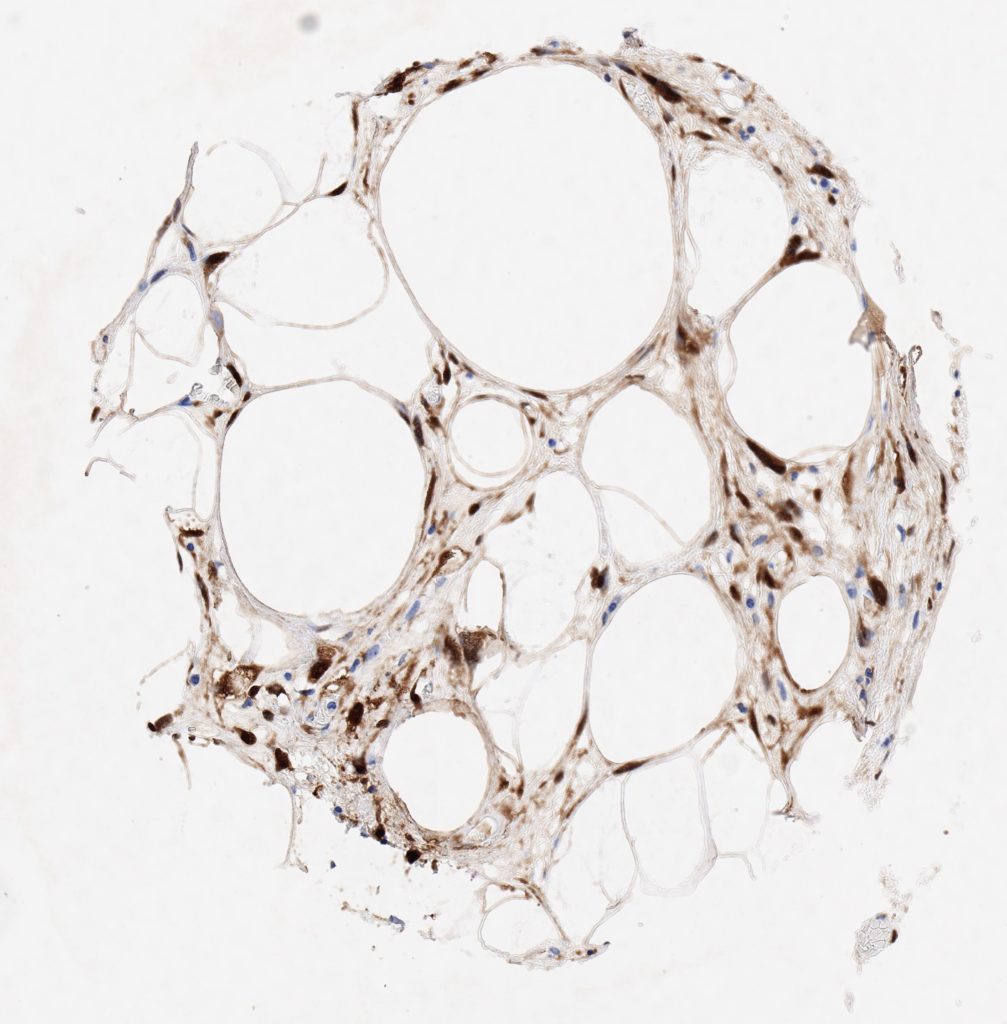
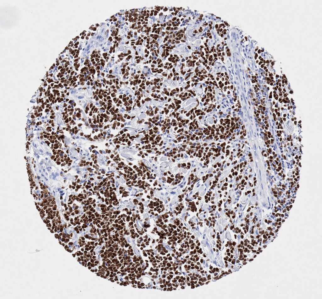
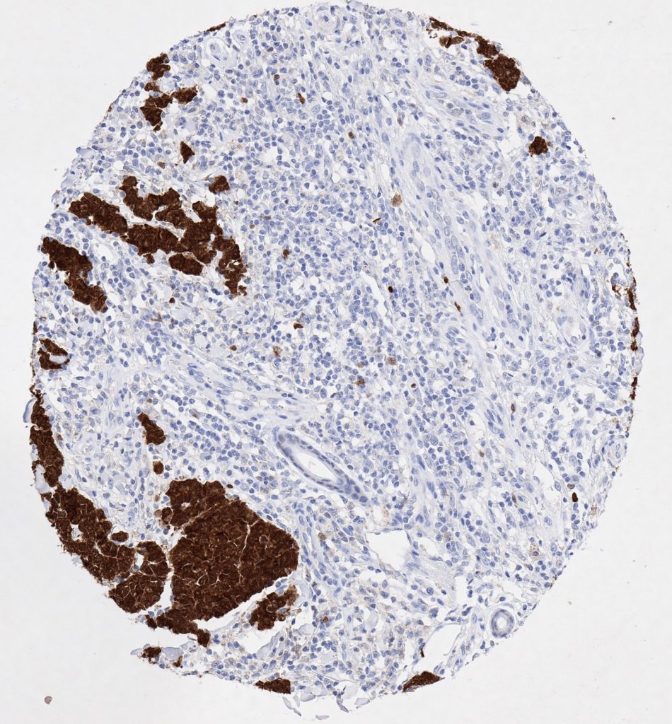
P16 spielt eine wichtige Rolle bei der Regulation des Zellzyklus. Es ist das wichtigste Mitglied der Ink4-Familie der Inhibitoren der cyclinabhängigen Kinase (CDK). Die Bindung von p16 hemmt die Bildung eines aktiven CDK4/6-Komplexes und die nachfolgende Phosphorylierung des Retinoblastom (Rb)-Proteins.  Da die Phospohorylierung des Rb-Proteins ein kritischer Schritt für das Fortschreiten des Zellzyklus von der G1- zur S-Phase ist, führt die p16-Bindung an die vorgeschaltete Kinase zum Stillstand des Zellzyklus. Folglich ist p16 ein negativer Regulator der Zellproliferation und damit ein starker Tumorsuppressor.

Ca. 50% aller menschlichen Krebsarten zeigen eine Inaktivierung von p16, darunter Kopf- und Halskrebs, Ösophagus-, Gallenwegs-, Leber-, Lungen-, Blasen-, Kolon- und Brustkrebs, Leukämie, Lymphome und Glioblastome. Darüber hinaus wurde neben der Senkung der p16-Regulation bei Krebs auch eine p16-Überexpression bei HPV-(humanes Papillomavirus)-verwandten Tumoren, Gebärmutterhalskrebs und Plattenepithelkarzinomen des Kopfes und Halses beobachtet. Der p16-Rb-Signalweg ist ein Ziel für virale Onkoproteine. Das E7-Onkoprotein aus HPV inaktiviert Rb. Die p16-Überexpression in HPV-assoziierten Tumoren ist somit Ausdruck einer Dysregulation des Zellzyklus durch einen erfolglosen Versuch, die Zellproliferation zu stoppen.

p16 ist ein wichtiger immunhistochemischer (IHC) Marker in der gynäkologischen Pathologie.

Antibody Productdetails

Menü