p16

370,00 plus VAT

#DIA-P16-OD
Anti-p16 (Hu) aus Maus (Klon JAP16) – 100 µl
Kategorie:

Beschreibung

Prod.Nr. DIA-P16-OD
Spezifität p16
Speziesreaktivität Human
Speziesreaktivität Mouse
Isotyp IgG2b
Klon JAP16
Anwendung
Immunohistochemistry (IHC),
Formalin-fixed Paraffin-embedded sections (FFPE)
Western Blot
Konjugation unconjugated
Verdünnung IHC 1:100-1:200
Format
Liquid
Antibody purified (from culture supernatant)
Anzahl 100µl
Control tissue Cervival carcinoma
Format Tris pH 7.3-7.7, with 1% BSA, <0.1% NaN3
Anwendungshinweis Research Use Only
Hersteller ONCOdianova
p16 IHC-Gallery
IHC-figures and legends on p16 detection in different tumors.

Reaktivität

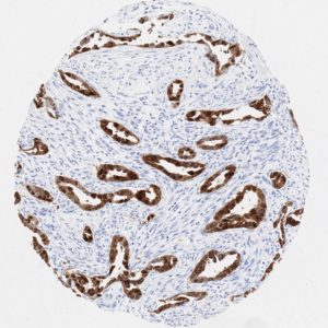
Clone JAP16 has been validated for the detection of p16 (INK4A) in routine formalin-fixed paraffin-embedded (FFPE) tissues. Anti-p16 clone JAP16 allows the identification of p16 in the tumors under pathological conditionsand provides valuable diagnostic information.

p16 plays an important role in cell cycle regulation. It is the principal member of the Ink4 family of cyclin-dependent kinase (CDK) inhibitors. Binding of p16 inhibits formation of an active CDK4/6 complex and subsequent phosphorylation of retinoblastoma (Rb) protein.  Since phospohorylation of Rb protein is a critical step for cell cycle progression from G1 to S phase, p16-binding to the upstream kinase leads to cell cycle arrest. Consequently, p16 is a negative regulator of cell proliferation and thus, a strong tumor suppressor.

Approx. 50% of all human cancers show p16 inactivation, these include head and neck, esophagus, biliary tract, liver, lung, bladder, colon and breast carcinomas; leukemia; lymphomas; and glioblastomas. Moreover, besides downregulation of p16 in cancer, p16 overexpression has been observed in HPV (human papilloma virus)-related tumors, cervical cancer and head and neck squamous carcinomas. The p16-Rb pathway is a target for viral oncoproteins. The E7 oncoprotein from HPV inactivates Rb. Thus, p16 overexpression in HPV-related tumors reflects cell cycle dysregulation by an unsuccessfull attempt to stop cell proliferation.

p16 is an important immunohistochemical (IHC) marker in gynecologic pathology.

IHC Protokoll

Staining protocols for anti-human p16 antibody clone JAP16
Cat.No.: DIA-P16-OD
Isotype: Mouse IgG2b
Specificity: Human p16
Physical State: Liquid
DAKO – autostainer Link 48
  • Pretreatment buffer: 15 min. / 95°C / pH9
  • Incubation primary antibody: 30 min. / RT (Dilution: 1:350)
  • Link: Flex-Mouse, 15 min. / RT
  • HRP (polymer): 20 min. / RT
BOND – Leica Bond RX
  • Pretreatment buffer: 20 min / 100°C / pH9
  • Incubation primary antibody: 15 min /RT (Dilution 1:350)
  • Post Primary: 8 min / RT
  • HRP (Polymer): 20 min / RT
Manual stain with autoclave
  • Pretreatment buffer: 121°C / 5 min / pH7,8
  • Incubation primary antibody: 60 min / 37°C (Dilution: 1:150)
  • Envision HRP rabbit/mouse: 30 min / 37°C

Referenzen

References

  1. Alcorta DA et al. (1996). Involvement of the cyclin-dependent kinase inhibitor p16 (INK4a) in replicative senescence of normal human fibroblasts. Proc Natl Acad Sci USA 93: 13742–13747.
  2. Serrano M. (1997). The tumor suppressor protein p16INK4a. Exp Cell Res 237: 7–13.
  3. Milde-Langosch K et al. (2001). Overexpression of the p16 cell cycle inhibitor in breast cancer is associated with a more malignant phenotype. Breast Cancer Res Treat 67: 61–70.
  4. Garcia V et al. (2004). Overexpression of p16INK4a correlates with high expression of p73 in breast carcinomas. Mutat Res 554: 215–221.
  5. Ohtani N et al. (2003). Epstein-Barr virus LMP1 blocks p16INK4a-RB pathway by promoting nuclear export of E2F4/5. J Cell Biol 162: 173–183
  6. Pei XH, Xiong Y. (2005). Biochemical and cellular mechanisms of mammalian CDK inhibitors: a few unresolved issues. Oncogene 24: 2787–2795
  7. Ivanova TA et al. (2007). Up-regulation of expression and lack of 5′ CpG island hypermethylation of p16 INK4a in HPV-positive cervical carcinomas. BMC Cancer 7: 47
  8. Reuschenbach M et al. (2008). Characterization of humoral immune responses against p16, p53, HPV16 E6 and HPV16 E7 in patients with HPV-associated cancers. Int J Cancer 123: 2626–2631.
  9. Paulson TG et al. (2008). p16 mutation spectrum in the premalignant condition Barrett’s esophagus. PLoS One 3: e3809
  10. Mulvany NJ et al. (2008). Diagnostic utility of p16INK4a: a reappraisal of its use in cervical biopsies. Pathology 40: 335–344.
  11. Lam AK et al. (2008). p16 expression in colorectal adenocarcinoma: marker of aggressiveness and morphological types. Pathology 40: 580–585.
Menü