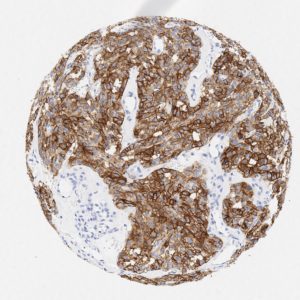
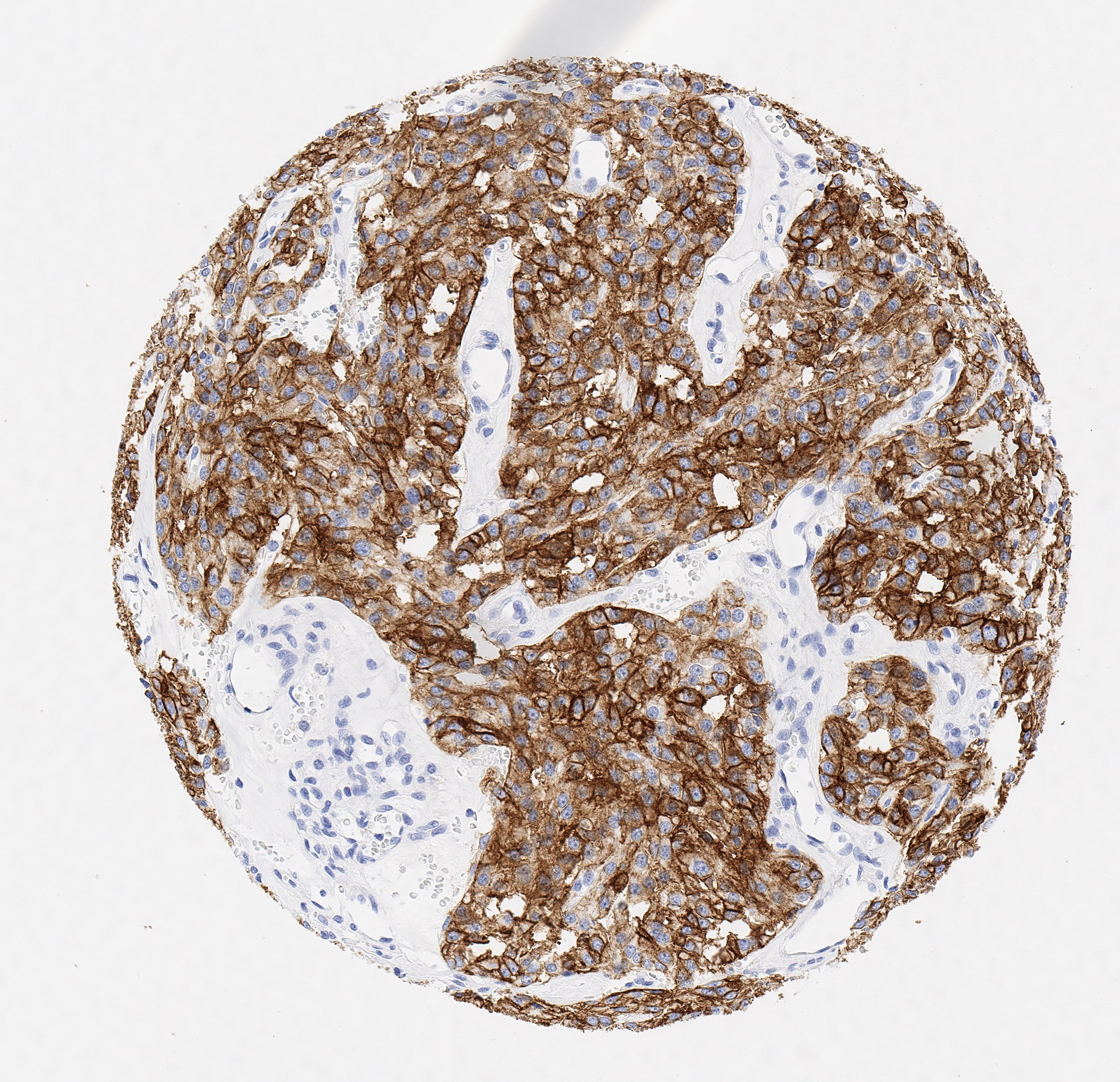
PD-L1

488,00 plus VAT

#DIA-PDL1-OD
Anti-PD-L1 (Hu) aus Maus (Klon JAL1) – 100 µl
Kategorie:

Beschreibung

Prod.Nr. DIA-PDL1-OD
Spezifität PD-L1
Speziesreaktivität Human
Isotyp Rabbit IgG
Klon JAL1
Anwendung
Immunohistochemistry (IHC),
Formalin-fixed Paraffin-embedded sections (FFPE)
Western Blot
Konjugation unconjugated
Verdünnung IHC 1:100-1:200
Format
Liquid
Antibody purified (from culture supernatant)
Anzahl 100µl
Control tissue Tonsil
Format Tris pH 7.3-7.7, with 1% BSA, <0.1% NaN3
Anwendungshinweis Research Use Only
Hersteller ONCOdianova
PD-L1 IHC-Gallery
IHC-figures and legends on p16 detection in different tumors.

Reaktivität

Clone JAL1 has been developed specifically for routine immunohistochemical (IHC) detection of PD-L1 in formalin-fixed paraffin-embedded tissue specimen. Moreover, JAL1 has been validated for the identification of PD-L1-positive macrophages and tumor tissues under pathological conditions.

PD-L1, also known as CD274 or B7 homolog 1 (B7-H1) is highly expressed in the heart, skeletal muscle, placenta and lung and weakly expressed in the thymus, spleen, kidney and liver. PD-L1 is expressed on macrophages and activated T- and B-cells, dendritic cells, keratinocytes and monocytes.

Moreover, several human cancer cells express PD-L1 at high levels. Binding of PD-L1 with its receptor PD-1 on T cells inhibits t cell proliferation and the production of cytokines, such as interleukin (IL)-2. It has been shown that PD-L1 helps tumor cells to evade anti-tumor immunity and blockade of PD-L1 reduces the growth of tumors in the presence of immune cells.

PD-L1 is commonly over expressed on tumor cells or on non-transformed cells in the tumor microenvironment2. PD-L1 expressed on the tumor cells binds to PD-1 receptors on the activated T cells, which leads to the inhibition of the cytotoxic T cells. These deactivated T cells remain inhibited in the tumor microenvironment.

PD-1 and PD-L1/PD-L2 belong to the family of immune checkpoint proteins that act as co-inhibitory factors, which can halt or limit the development of the T cell response.

Referenzen

References.

  1. O’Malley DP et al. Immunohistochemical detection of PD-L1 among diverse human neoplasms in a reference laboratory: observations based upon 62,896 cases. Modern Pathology (2019) 32:929–942
  2. Büttner R et al. Programmed death-ligand 1 immunohistochemistry testing: a review of analytical assays and clinical implementation in non-small-cell lung cancer. J Clin Oncol. (2017) 35:3867–3876
  3. Rimm DL et al. A prospective, multi-institutional, pathologist-based assessment of 4 immunohistochemistry assays for PD-L1 expression in non-small cell lung cancer. JAMA Oncol. (2017) 3:1051–1058
  4. Milne CP et al. Complementary versus companion diagnostics: apples and oranges? Biomark Med. (2015) 9:25–34
  5. Ott PA et al. CTLA-4 and PD-1/PD-L1 blockade: new immunotherapeutic modalities with durable clinical benefit in melanoma patients. Clin Cancer Res. (2013) 19:5300–5309
  6. Pardoll DM. The blockade of immune checkpoints in cancer immunotherapy. Nat Rev Cancer. (2012) 12:252–264.
  7. Yokosuka T et al. Programmed cell death 1 forms negative costimulatory microclusters that directly inhibit T cell receptor signaling by recruiting phosphatase SHP2. J Exp Med. (2012) 209:1201–1217
Menü